
Antibody and Dye Based Assays


Biotium Cytokeratin 8(H1), CF488A conjugate, 0.1mg/mL
Cytokeratin 8 (CK8) belongs to the type II (or B or basic) subfamily of high molecular weight cytokeratins and exists in combination with cytokeratin 18 (CK18). CK8 is primarily found in the non-squamous epithelia and is present in majority of adenocarcinomas and ductal carcinomas. It is absent in squamous cell carcinomas. Hepatocellular carcinomas are defined by the use of antibodies that recognize only cytokeratin 8 and 18. CK8 exists on several types of normal and neoplastic epithelia, including many ductal and glandular epithelia such as colon, stomach, small intestine, trachea, and esophagus as well as in transitional epithelium. Anti-CK8 does not react with skeletal muscle or nerve cells. Epithelioid sarcoma, chordoma, and adamantinoma show strong positivity corresponding to that of simple epithelia (with antibodies against CK8, CK18 and CK19). Reportedly, anti-CK8 is useful for the differentiation of lobular (ring-like, perinuclear) from ductal (peripheral-predominant)

Biotium Cytokeratin, LMW(AE-1), CF488A conjugate, 0.1mg/mL
This MAb recognizes the 56.5 kDa (CK10); 50 kDa (CK14); 50 kDa (CK15); 48 kDa (CK16); 40 kDa (CK19) keratins of the acidic (Type I or LMW) subfamily. Twenty human keratins are resolved with two-dimensional gel electrophoresis into acidic (pI 6.0) subfamilies. The acidic keratins have molecular weights (MW) of 56.5, 55, 51, 50, 50', 48, 46, 45, and 40 kDa. MAb AE3 recognizes the 65-67, 64, 59, 58, 56, and 52 kDa keratins of basic subfamily. Many studies have shown the usefulness of keratins as markers in cancer research and tumor diagnosis. AE1/AE3 is a broad spectrum anti pan-keratin antibody cocktail, which differentiates epithelial tumors from non-epithelial tumors e.g. squamous vs. adenocarcinoma of the lung, liver carcinoma, breast cancer, and esophageal cancer.Primary antibodies are available purified, or with a selection of fluorescent CF Dyes and other labels. CF Dyes offer exceptional brightness and photostability. Note: Conjugates of blue fluorescent dyes like CF
Biotium CD53(63-5A3), CF488A conjugate, 0.1mg/mL
Recognizes a protein of 33-55 kDa, identified as CD53 (Workshop V; Code CD53.1). CD53 is expressed on monocytes, and macrophages, granulocytes, dendritic cells, osteoblasts and osteoclasts, NK cells, and on T- and B-cells from every stage of differentiation but is absent from platelets, erythrocytes, and non-haemopoietic cells. CD53 is a member of a family of tetraspan transmembrane proteins, including CD9, CD37, CD63, CD81, and CD82. It associates with integrins, MHC class II molecules, and a tyrosine phosphatase and plays a role in cellular activation as part of a signal transduction complex involving other membrane glycoproteins. Defects of CD53 expression on neutrophils appear to be related with recurrent infectious diseases. Cross-linking CD53 using CD53 antibodies led to cytoplasmic calcium fluxes in B cells, monocytes, and granulocytes and activation of the monocyte oxidative burst.Primary antibodies are available purified, or with a selection of fluorescent CF Dyes a
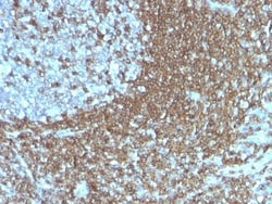
Biotium CD50 (ICAM-3)(186-2G9), CF488A conjugate, 0.1mg/mL
Recognizes the D1 domain of an N-glycosylated glycoprotein of 120 kDa with intra-chain disulfide bonds, identified as CD50 or ICAM-3 (Leucocyte Workshop VI). CD50 is the major ligand for LFA-1 (CD11a/CD18) and may have signaling role to increase adhesion. It is expressed on thymocytes and T lymphocytes and is resistant to treatment with phosphatidylinositol (PI) phospholipase C. This MAb is excellent for staining of formalin/paraffin tissues.Primary antibodies are available purified, or with a selection of fluorescent CF Dyes and other labels. CF Dyes offer exceptional brightness and photostability. Note: Conjugates of blue fluorescent dyes like CF405S and CF405M are not recommended for detecting low abundance targets, because blue dyes have lower fluorescence and can give higher non-specific background than other dye colors.
Biotium CD48(156-4H9), CF488A conjugate, 0.1mg/mL
Reacts with human CD48, a 45 kDa glycosyl phophatidyl-inositol (GPI)-anchored cell surface protein. CD48 is strongly expressed on lymphocytes and monocytes and weakly on granulocytes but is absent on platelets, fibroblasts, epithelium and endothelium. CD48 is one of marker for detecting the defects of GPI anchoring structure on the patients with paroxysmal nocturnal hemoglobulinuria (PNH) and serves as a low affinity ligand for CD2.Primary antibodies are available purified, or with a selection of fluorescent CF Dyes and other labels. CF Dyes offer exceptional brightness and photostability. Note: Conjugates of blue fluorescent dyes like CF405S and CF405M are not recommended for detecting low abundance targets, because blue dyes have lower fluorescence and can give higher non-specific background than other dye colors.
Electron Microscopy Sciences Bio-Pump Via-Cell Flow Indicator
A convenient, easy to use flow indicator used for calibration of the Zefon Bio-Pump and Bio-Pump Plus.

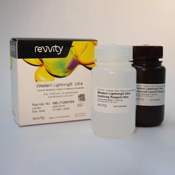
Revvity Health Sciences Inc Western Lightning Ultra, Chemiluminescent Substrate (110 mL)
Western Lightning Ultra, Chemiluminescent Substrate (110 mL)
Biotium Desmocollin-2/3 (7G6), CF488A conjugate, 0.1mg/mL
Desmosomes are intercellular adhering junctions that represent cell surface attachment sites for intermediate filament. The desmosome is subdivided into two regions. The plaque region lies adjacent to the plasma, and is believed to contain molecules that attach the intermediate filament cytoskeleton to the desmosome. The core region is composed of transmembrane glycoproteins that are thought to mediate cell-cell adhesion. Desmogleins and desmocollins are the main desmosomal transmembrane proteins. These desmosomal glycoproteins belong to the members of the cadherin family of adhesion molecules. Three different isoforms of both desmogleins and desmocollins have been identified, named as desmoglein 1-3 and desmocollins. Desmosomal cadherins showed differentiation-specific expression in the human epidermis, although the functional significance of this differential expression is not fully understood. Desmocollin-1 can be found in the upper layers. The expression of desmocollin-2
Biotium CD81 / TAPA-1 (rC81/3442), CF488A conjugate, 0.1mg/mL
This antibody recognizes a protein of 26 kDa, identified as CD81 (Workshop VI; Code CD81.1)). CD81 has a very broad cellular distribution, being expressed on T- and B-lymphocytes, NK cells, thymocytes, eosinophils, fibroblasts, epithelial and endothelial cells. Neutrophils, erythrocytes and platelets are negative, while monocytes are variably positive. CD81 is a member of a family of tetraspanin transmembrane proteins, including CD9, CD37, CD53, CD63, and CD82. It associates with CD19, CD21, Leu 13, and integrins on cell membrane and is involved in signal transduction in B lymphocyte development and cell adhesion. CD81 also acts as a receptor for the envelope protein E2 of chronic hepatitis C virus. Antibodies to CD81 have anti-proliferative effects on different lymphoid cell lines, particularly those derived from large cell lymphomas.Primary antibodies are available purified, or with a selection of fluorescent CF Dyes and other labels. CF Dyes offer exceptional brightness a
Biotium CD3e (T-Cell Marker)(OKT3), CF488A conjugate, 0.1mg/mL
Recognizes the epsilon-chain of CD3, which consists of five different polypeptide chains (designated as gamma, delta, epsilon, zeta, and eta) with MW ranging from 16-28 kDa. CD3, also known as T3, is a member of the immunoglobulin superfamily that plays a role in antigen recognition, signal transduction, and T cell activation. It is found on all mature T-lymphocytes, NK, T-cells, and some thymocytes, and is expressed, typically at high levels, on peripheral T cells and majority of T cell neoplasms. Thymocytes express CD3 at different level on the cell surface in the course of differentiation and CD3 is predominantly intracytoplasmic in cortical thymus.Anti-CD3 clone OKT3 has been reported to have potent immunosuppressive properties in vivo and has been proven effective in the treatment of renal, heart and liver allograft rejection. In the crystal structure of the antibody-receptor complex, clone OKT3 was found to bind a conformational epitope on CD3e dominated by a linear
Biotium Calnexin (Endoplasmic Reticulum Marker) (CANX/1541), Biotin conjugate, 0.1mg/mL
Calnexin is a type-I integral membrane protein of the endoplasmic reticulum (ER). It is a calcium-binding chaperone protein that interacts transiently with newly synthesized N-linked glycoproteins, facilitating protein folding and assembly. It may also play a central role in the quality control of protein folding by retaining incorrectly folded protein subunits within the ER for degradation.Primary antibodies are available purified, or with a selection of fluorescent CF Dyes and other labels. CF Dyes offer exceptional brightness and photostability. Note: Conjugates of blue fluorescent dyes like CF405S and CF405M are not recommended for detecting low abundance targets, because blue dyes have lower fluorescence and can give higher non-specific background than other dye colors.
Biotium Calnexin (Endoplasmic Reticulum Marker) (CANX/1541), CF568 conjugate, 0.1mg/mL
Calnexin is a type-I integral membrane protein of the endoplasmic reticulum (ER). It is a calcium-binding chaperone protein that interacts transiently with newly synthesized N-linked glycoproteins, facilitating protein folding and assembly. It may also play a central role in the quality control of protein folding by retaining incorrectly folded protein subunits within the ER for degradation.Primary antibodies are available purified, or with a selection of fluorescent CF Dyes and other labels. CF Dyes offer exceptional brightness and photostability. Note: Conjugates of blue fluorescent dyes like CF405S and CF405M are not recommended for detecting low abundance targets, because blue dyes have lower fluorescence and can give higher non-specific background than other dye colors.
Biotium CD3e (T-Cell Marker)(OKT3), CF488A conjugate, 0.1mg/mL
Recognizes the epsilon-chain of CD3, which consists of five different polypeptide chains (designated as gamma, delta, epsilon, zeta, and eta) with MW ranging from 16-28 kDa. CD3, also known as T3, is a member of the immunoglobulin superfamily that plays a role in antigen recognition, signal transduction, and T cell activation. It is found on all mature T-lymphocytes, NK, T-cells, and some thymocytes, and is expressed, typically at high levels, on peripheral T cells and majority of T cell neoplasms. Thymocytes express CD3 at different level on the cell surface in the course of differentiation and CD3 is predominantly intracytoplasmic in cortical thymus.Anti-CD3 clone OKT3 has been reported to have potent immunosuppressive properties in vivo and has been proven effective in the treatment of renal, heart and liver allograft rejection. In the crystal structure of the antibody-receptor complex, clone OKT3 was found to bind a conformational epitope on CD3e dominated by a linear

Revvity Health Sciences Inc HTRF Human MDA5 Aggregation Kit, 500 Assay Points
HTRF Human MDA5 Aggregation Kit, 500 Assay Points
Biotium CD28 (C28/1636), CF488A conjugate, 0.1mg/mL
This antibody recognizes a glycoprotein of 44-88 kDa, which is identified as CD28. It is the critical T-cell co-stimulatory receptor which provides to the cell the important second activation signal by binding CD80 and CD86 that are expressed by antigen presenting cells. Besides its co-stimulation role, CD28 functions in preventing T-cells from anergic hyporesponsive state or from undergoing premature apoptotic cell death. CD28 is also expressed on human fetal NK cells and some NK cell lines, whereas on murine NK cells the CD28 expression is much broader.Primary antibodies are available purified, or with a selection of fluorescent CF Dyes and other labels. CF Dyes offer exceptional brightness and photostability. Note: Conjugates of blue fluorescent dyes like CF405S and CF405M are not recommended for detecting low abundance targets, because blue dyes have lower fluorescence and can give higher non-specific background than other dye colors.