Learn More
Invitrogen™ CD223 (LAG-3) Monoclonal Antibody (3DS223H), Brilliant Violet™ 421, eBioscience™, Invitrogen™


Description
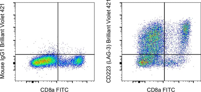
This 3DS223H mAb recognizes human CD223 also known as Lymphocyte Activation Gene 3 (LAG-3). LAG-3 is a 70-kDa surface glycoprotein belonging to the Ig superfamily with homology to CD4. LAG-3 binds to MHC class II with higher affinity than CD4 and is thought to be involved in the negative regulation of T cell activation and homeostatic proliferation. Surface expression of LAG-3 has been reported on activated T cells (including regulatory T cells) and NK cells. CD8+ T cells usually express LAG-3 at significantly higher levels than CD4+ T cells. Coexpression of LAG-3 and CD49b has been proposed to identify human and mouse Type 1 regulatory T cells (Tr1 cells). This 3DS223H antibody will recognize a formaldehyde-fixed epitope. Applications Tested: This 3DS223H antibody has been pre-diluted and tested by flow cytometric analysis of stimulated normal human peripheral blood cells. BV 421 is a dye that emits at 423nm and is intended for use on cytometers equipped with a violet (405nm) laser. When using two or more Super Bright, BV, BUV, or other polymer dye-conjugated antibodies in a staining panel, it is recommended to use Super Bright Complete Staining Buffer (SB-4401-42) or Brilliant Stain Buffer (00-4409-75) to minimize any non-specific polymer interactions. Excitation: 407nm; Emission: 423nm; Laser: Violet Laser. BV is a trademark or registered trademark of Becton, Dickinson and Company or its affiliates, and is used under license. Powered by Sirigen.
Specifications
Specifications
| Antigen | CD223 (LAG-3) |
| Applications | Flow Cytometry |
| Classification | Monoclonal |
| Clone | 3DS223H |
| Concentration | 5 μL/Test |
| Conjugate | Brilliant Violet 421 |
| Formulation | PBS with BSA and 0.09% sodium azide; pH 7.2 |
| Gene | LAG3 |
| Gene Accession No. | P18627 |
| Gene Alias | Activation-induced cytidine deaminase-linked autoimmunity protein; Aida; CD223; FDC; LAG3; LAG-3; Ly66; lymphocyte activating 3; lymphocyte activation gene 3 protein; lymphocyte-activation gene 3; Secreted lymphocyte activation gene 3 protein; sLAG 3; sLAG3; sLAG-3; soluble LAG 3lymphocyte activating 3; soluble LAG3 |
| Show More |
By clicking Submit, you acknowledge that you may be contacted by Fisher Scientific in regards to the feedback you have provided in this form. We will not share your information for any other purposes. All contact information provided shall also be maintained in accordance with our Privacy Policy.