Learn More
Invitrogen™ IDO Monoclonal Antibody (eyedio), Brilliant Ultra Violet™ 615, eBioscience™

Description
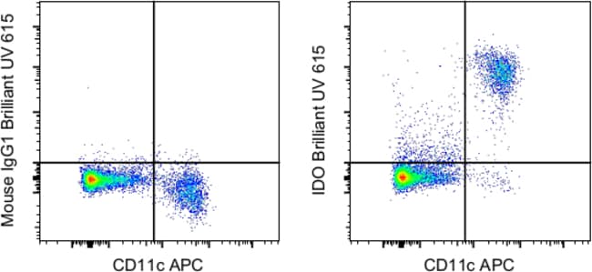
Description This eyedio monoclonal antibody reacts with human indoleamine-2,3-dioxygenase (IDO, INDO, IDO1), an intracellular enzyme that catalyzes the degradation of tryptophan to kynurenines. IDO is expressed in a wide variety of tissues and cells, including macrophages, plasmacytoid dendritic cells, and several cell lines. IDO can be induced in many different cell types by IFN gamma or other inflammatory stimuli. Expression of IDO in antigen presenting cells and tumors is thought to mediate immune suppression through depletion of this essential amino acid and/or by the creation of tryptophan metabolites that cause apoptosis of T cells and induction of regulatory T cells. Applications Tested This eyedio antibody has been pre-diluted and tested by intracellular staining followed by flow cytometric analysis of normal human peripheral blood cells using the Intracellular Fixation & Permeabilization Buffer Set (Product # 88-8824-00) and protocol. Please refer to 'Staining Intracellular Antigens for Flow Cytometry, Protocol A: Two step protocol for intracellular (cytoplasmic) proteins' located at Flow Protocols>. This may be used at 5 μL (0.25 μg) per test. A test is defined as the amount (μg) of antibody that will stain a cell sample in a final volume of 100 μL. Cell number should be determined empirically but can range from 10^5 to 10^8 cells/test. Blocking Buffers When using two or mor...
Specifications
Specifications
| Antigen | IDO |
| Applications | Flow Cytometry |
| Classification | Monoclonal |
| Clone | eyedio |
| Concentration | 5 μL/Test |
| Conjugate | Brilliant Ultraviolet 615 |
| Formulation | PBS with BSA and 0.09% sodium azide; pH 7.2 |
| Gene | Ido1 |
| Gene Accession No. | P14902 |
| Gene Alias | EC 1.13.11.52; IDO; IDO1; IDO-1; INDO; indolamine 2,3 dioxygenase; indole 2,3-dioxygenase; indoleamine; indoleamine 2,3-dioxygenase 1; indoleamine 23-dioxygenase; Indoleamine-2; indoleamine-pyrrole 2,3 dioxygenase; indoleamine-pyrrole 2,3-dioxygenase |
| Show More |
By clicking Submit, you acknowledge that you may be contacted by Fisher Scientific in regards to the feedback you have provided in this form. We will not share your information for any other purposes. All contact information provided shall also be maintained in accordance with our Privacy Policy.