Promotional price valid on web orders only. Your contract pricing may differ. Interested in signing up for a dedicated account number?
Learn More
Learn More

Description
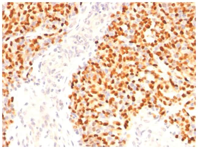
MyoD Monoclonal specifically detects MyoD in Human, Mouse, Rat, Chicken samples. It is validated for Western Blot, Flow Cytometry, Immunohistochemistry, Immunocytochemistry/Immunofluorescence, Immunohistochemistry-Paraffin, Flow (Intracellular).
Specifications
Specifications
| Antigen | MyoD |
| Applications | Western Blot, Flow Cytometry, Immunocytochemistry, Immunofluorescence, Immunoprecipitation, Immunohistochemistry (Paraffin) |
| Classification | Monoclonal |
| Clone | SPM427 |
| Conjugate | Unconjugated |
| Dilution | Western Blot 0.25-0.5ug/ml, Flow Cytometry 0.5-1ug/million cells, Immunocytochemistry/Immunofluorescence 0.5-1ug/ml, Immunoprecipitation 0.5-1ug/500ug protein lysate, Immunohistochemistry-Paraffin 0.5-1.0ug/ml, Immunohistochemistry-Frozen 0.5-1.0ug/ml |
| Formulation | PBS with 0.05% BSA. with 0.05% Sodium Azide |
| Gene Accession No. | P15172 |
| Gene Alias | bHLHc1BHLHC1, Class C basic helix-loop-helix protein 1, MYF3Myf-3, MYODmyoblast determination protein 1, myogenic differentiation 1, Myogenic factor 3myf-3, PUM |
| Gene Symbols | MYOD1 |
| Show More |
Product Title
By clicking Submit, you acknowledge that you may be contacted by Fisher Scientific in regards to the feedback you have provided in this form. We will not share your information for any other purposes. All contact information provided shall also be maintained in accordance with our Privacy Policy.
Spot an opportunity for improvement?