Promotional price valid on web orders only. Your contract pricing may differ. Interested in signing up for a dedicated account number?
Learn More

PD-1 Antibody (PDCD1/922), Novus Biologicals™
SDP

Catalog No. NBP24439801 Shop All R&D Systems Products
Change view
Click to view available options
Quantity:
0.02 mg
0.1 mg
0.2 mg
3 product options available for selection
Product selection table with 3 available options. Use arrow keys to navigate and Enter or Space to select.
Catalog No. Quantity
NBP24439801 0.1 mg
NBP24439800 0.02 mg
NBP24439802 0.2 mg
Use arrow keys to navigate between rows. Press Enter or Space to select a product option. 3 options available.
3 options
Catalog No. NBP24439801 Supplier Novus Biologicals Supplier No. NBP2443980.1MG
Only null left
Add to Cart
Add to Cart

Mouse Monoclonal Antibody

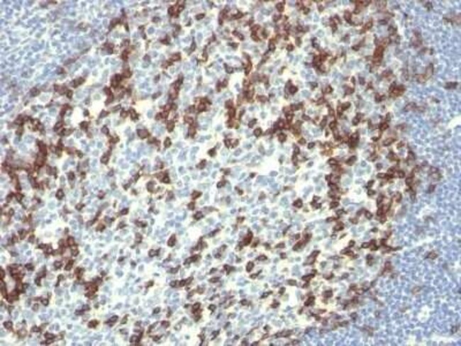
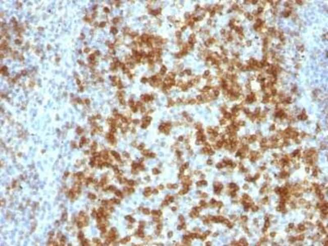
Ensure accurate, reproducible results in Flow Cytometry, Immunohistochemistry (Paraffin), Immunofluorescence

PD-1 Monoclonal specifically detects PD-1 in Human samples. It is validated for Flow Cytometry, Immunohistochemistry, Immunocytochemistry/Immunofluorescence, Immunohistochemistry-Paraffin, Immunofluorescence.

Specifications

Antigen PD-1
Applications Flow Cytometry, Immunohistochemistry (Paraffin), Immunofluorescence
Classification Monoclonal
Clone PDCD1/922
Concentration 0.2mg/mL
Conjugate Unconjugated
Dilution Flow Cytometry 0.5 - 1 ug/million cells in 0.1 ml, Immunohistochemistry-Paraffin 0.5 - 1.0 ug/ml, Immunofluorescence 1 - 2 ug/ml
Formulation 10mM PBS and 0.05% BSA with 0.05% Sodium Azide
Gene Accession No. Q15116
Gene Alias CD279, CD279 antigen, hPD-1, PD1hPD-l, programmed cell death 1, programmed cell death protein 1, Protein PD-1, SLEB2
Gene Symbols PDCD1
Host Species Mouse
Immunogen Recombinant full-length human PDCD1 protein
Molecular Weight of Antigen 55 kDa
Purification Method Protein A or G purified
Quantity 0.1 mg
Regulatory Status RUO
Research Discipline Apoptosis, Stem Cell Markers
Primary or Secondary Primary
Gene ID (Entrez) 5133
Test Specificity PDCD-1 (programmed cell death-1 protein), also designated CD279, is a type I transmembrane receptor and a member of the immunoglobin gene superfamily. It is expressed on activated T-cells, B-cells, and myeloid cells. Anti-PDCD-1 is a marker of angioimmunoblastic lymphoma and suggests a unique cell of origin for this neoplasm. Unlike CD10 and BCL6, PDCD-1 is expressed by few B-cells, so anti-PDCD-1 may be a more specific and useful diagnostic marker in angioimmunoblastic lymphoma. In addition, PDCD-1 expression provides evidence that angioimmunoblastic lymphoma is a neoplasm derived from germinal center-associated T-cells.
Target Species Human
Content And Storage Store at 4C.
Form Purified
Isotype IgG1 κ
Show More Show Less

For Research Use Only

Product Title
Select an issue

By clicking Submit, you acknowledge that you may be contacted by Fisher Scientific in regards to the feedback you have provided in this form. We will not share your information for any other purposes. All contact information provided shall also be maintained in accordance with our Privacy Policy.