Promotional price valid on web orders only. Your contract pricing may differ. Interested in signing up for a dedicated account number?
Learn More
Learn More
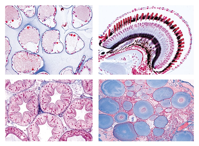
Johannes Lieder The Crayfish (Astacus)

Shop All Johannes Lieder GmbH & Co. KG Products
Click to view available options
Mounting Type:
Cross and Longitudinal Section
Description
- 5700 The Crayfish (Astacus fluviatilis)
- 12 Microscope Slides
- Crayfish, gills t.s., epithelium and vessels
- Crayfish, striated muscle l.s., showing striations very clearly
- Crayfish, antenna (decalcified) t.s., showing the chitinous skeleton
- Crayfish, compound eye l.s.
- Crayfish, cerebral ganglion t.s., nerve cells and fibers
- Crayfish, blood smear, with blood cells
- Crayfish, green gland t.s., an excretory organ
- Crayfish, stomach t.s., internal chitinous layer
- Crayfish, intestine t.s., folds of mucous membrane
- Crayfish, liver t.s., glandular tubules for reabsorption of food
- Crayfish, ovary t.s., development of ova
- Crayfish, testis t.s., spermatogenesis, cell division stages
Specifications
Specifications
| Classification | Zoology |
| Material | Elastin Stained Glass |
| Mounting Type | Cross and Longitudinal Section |
| Type | Microscope Slides |
| Slide Type | Prepared Slide |
Product Content Correction
Your input is important to us. Please complete this form to provide feedback related to the content on this product.
Product Title
Spot an opportunity for improvement?Share a Content Correction