
Incubator Accessories

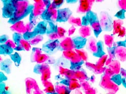
Diagnostic Biosystems PAPANICOLAOU RED STAIN KIT Papanicolaou Red Stain Kit
The Papanicolaou (PAP) Stain Kit is designed to differentiate between a variety of cells in vaginal smears for detection of vaginal, uterine and cervical cancer.


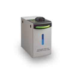
Caron Products And Services Universal Condensate Recirculating System; 230V, 50/60Hz
These recirculators act as a water delivery system for use with any Caron humidity controlled chamber.


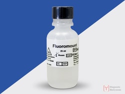
Diagnostic Biosystems FLUOROMOUNT/PLUS Fluoromount/Plus™
Fluoromount/Plus™ is an innovative, aqueous-based mounting medium designed especially for the permanent preservation of fluorescent specimens. Fluoromount/Plus™is to be used with tissue sections stained with a multitude of fluorescent dyes. This product is compatible with FITC, phycoerythrin, phycocyanin and allophycocyanin. It can also be used with other fluorescent markers such as Texas Red and rhodamine.\n\nMounting tissue specimens stained with fluorescent dyes.
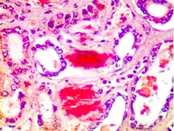
Diagnostic Biosystems AMYLOID Amyloid Stain Kit (Congo Red)
The Amyloid Stain Kit (Congo Red) is intended for use in the histological visualization of amyloid in tissue sections. Examination under a polarizing microscope results in green birefringence of amyloid.
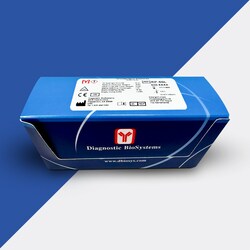
Diagnostic Biosystems PEROXIDASE Universal HRP Immunostaining Kit (For Mouse and Rabbit Primary Antibodies)
This product is intended for qualitative immunohistochemistry with normal and neoplastic formalin-fixed, paraffin-embedded tissue sections, tobe viewed by light microscopy. Clinical interpretation of staining results should be accompanied by histological studies with proper controls. Patients’ clinical histories and other relevant diagnostic tests should be utilized by a qualified person (s) when evaluating and interpreting results. This kit is designed to label specific primary antibodies immunohistochemically on tissue sections. The immunohistochemical protocol defined in this brochure is just a guideline. Depending upon tissue fixation conditions, the primary antibody employed, and the user's experience, we encourage individual laboratory to optimize its own protocol. These reagents were tested and quality controlled using tissue sections, however, they can also be optimized for cell smears and cytospin preparations. Sufficient reagents are provided to run 50 to 100 tests.
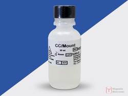
Diagnostic Biosystems CC/MOUNT CC/Mount™
CC/Mount is an aqueous mounting medium with very high refractive index. When applied to the stained tissue sections, specimens can be permanently mounted without chromogens fading. Due to the superior refractive index of CC/Mount, tissues mounted in this medium look like dehydrated specimens. No coverslipping is required with CC/Mount. However, if coverslipping is desired, dried CC/Mount can be post-mounted by using an organic based mounting medium. Advantages of CC/Mount are numerous: no coverslip, no exposure to the organic fumes, permanent storage of slides and high resolution of tissue specimens. CC/Mount is compatible with chromogens lik AEC, DAB, Fast Red, BCIP/NBT, BCIP/INT and fluorescent dyes like FITC and phycobiliproteins. The high pH of CC/Mount ensures increased stability of fluorescence.\n\nThis product is designed for permanent mounting of tissue specimens stained with peroxidase and alkaline phosphatase-based systems and various fluorescent dyes.
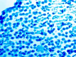
Diagnostic Biosystems LUXOL FAST BLUE Luxol Fast Blue
The Luxol Fast Blue Stain Kit is designed for staining myelin/myelinated axons and Nissil substance on formalin fixed, paraffin-embedded tissue. This product is used for identifying the basic neuronal structure in brain or spinal cord sections.
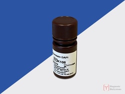
Diagnostic Biosystems DAPI
DAPI or 4′,6-diamidino-2-phenylindole, is a fluorescent stain that binds strongly to adenine–thymine-rich regions in DNA. It is used extensively in fluorescence microscopy. As DAPI can pass through an intact cell membrane, it can be used to stain both live and fixed cells.\n\nFeatures\n• This DAPI solution is optimized for use on formalin-fixed, paraffin-embedded tissue sections.\n• The DAPI solution contains an anti-freeze agent so the reagent can be stored at -20 °C without freezing.\n• For convenience the DAPI solution can be removed from the freezer and used immediately without the necessity of thawing.\n• There is no need to aliquot and freeze small portions.\n\nKnown Applications\n• Fluorescence In Situ Hybridization\n• Immunofluorescence with fluorescently labeled antibodies or probes.\n• Fluorescent staining methods.

Diagnostic Biosystems CD138 CD138 (EP201)
CD138, also known as Syndecan-1, is a member of the transmembrane heparan sulfate proteoglycan family, acts as an extracellular matrix receptor and is involved in many cellular functions, including cell-cell adhesion and cell-matrix adhesion. CD138 expression is found in both hematopoietic and non-hematopoietic cells. In the hematopoietic system, CD138 labels plasma cells. It is an excellent marker for plasmacytic differentiation within the spectrum of hematologic malignancy. Among non-hematolymphoid cells, CD138 reactivity is observed in many types of epithelial cells and stoma cells in both normal and tumor tissues.
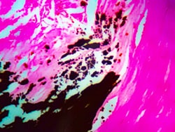
Diagnostic Biosystems VON KOSSA Von Kossa
The Calcium Stain Kit (Modified Von Kossa) is intended for use in the histological visualization of calcium deposits in paraffin sections.
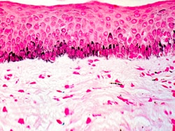
Diagnostic Biosystems FONTANA-MASSON Fontana-Masson
The Fontana-Masson Stain Kit is intended for use in the histological visualization of Argentaffin cells and Melanin in paraffin or frozen sections.
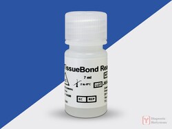
Diagnostic Biosystems TISSUE BOND Tissue Bond™ Reagent
TissueBond reacts with silica glass to change its surface chemistry so that it may bond either ionically or covalently with aldehyde and ketone groups in tissue sections.\n\nProper tissue adhesion to glass slides is a critical step in immunohistochemistry. Good immunostaining may require harsh pretreatment of tissues for antigen retrieval (e.g. proteolytic enzyme digestion, boiling in high pH buffer, treatment with detergents or saponin). Improper adhesion of tissue sections can lead to poor quality staining or even loss of tissue sections. White glue, albumen, chrome-gelatin, and poly-L-lysine increase background staining. There is a need for a tissue adhesive that can irreversibly bind tissue sections to glass without creating background staining. Glass treated with TissueBond can be used for both paraffin and frozen tissue sections.