
Incubator Accessories
Diagnostic Biosystems KAPPA LIGHT CHAIN Kappa Light Chain
This antibody reacts with free as well as bound kappa light chains. Contaminating antibodies have been removed by solid phase adsorption.


Nuaire Inc CO2 INCUBATOR TUBING PER FOOT

This item has a minimum qty of 13 per supplier requirements.

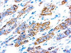
Diagnostic Biosystems HER2/NEU C-ERBB-2 c-erbB-2 Oncoprotein (SP3)
This antibody recognizes a c-erbB-2 protein, which is a receptor tyrosine kinase of the c-erbB family. It is closely related in structure to the epidermal growth factor receptor. c-erbB-2 oncoprotein is detectable in a subset of breast and other adenocarcinomas, as well astransitional cell carcinomas. In the case of breast cancer, expression determined byimmunohistochemistry has been shown to be associated with poor prognosis.
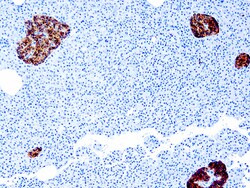
Diagnostic Biosystems CHROMOGRANIN A Chromogranin A (SP12)
This antibody recognizes Chromogranin A. Chromogranin A (a protein of 439 amino acid which is encoded on chromosome 14) is present in neuroendocrine cells throughout the body, including the neuroendocrine cells of the large and small intestine, adrenal medulla and pancreatic islets. It is an excellent marker for carcinoid tumors, pheochromocytomas, paragangliomas, and other neuroendocrine tumors. Coexpression of chromogranin A and neuron specific enolase (NSE) is common in neuroendocrine neoplasms.
Diagnostic Biosystems MLH-1 MLH-1
The G168-15 antibody recognizes human and mouse MLH1 (80-85kDa). The repair of mismatch DNA is essential to maintaining the integrity of genetic information over time. An alteration of microsatellite repeats is the result of slippage owing to strand misalignment during DNA replication and is referred to as microsatellite instability (MSI). These defects in DNA repair pathways have been related to human carcinogenesis. The importance of mismatch repair genes became apparent with the identification of the genetic basis for hereditary nonpolyposis colon cancer (HNPC). MSH-2 isinvolved in the initial cognition of mismatch nucleotides during the replication mismatch repair process. It is thought that after MSH2 binds to a mismatched DNA duplex it isjoined by a heterodimer of MLH1 and PMSH, which together help facilitate the later steps in mismatch repair.
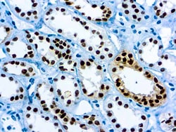
Diagnostic Biosystems PAX-8 PAX-8
PAX 8 is expressed in a high percentage of renal cell carcinomas and ovarian cancers. This mouse monoclonal PAX 8 antibody [4H7B3] has been designed to target restricted epitopes, and exhibits higher specificity and provides sharper staining than the PAX 8 rabbit polyclonal antibody. Unlike the polyclonal PAX 8, this mouse monoclonal antibody does not stain B-cells, and does not recognize epitopes of pancreatic origin and neuroendocrine cells in stomach and colon; thus providing superior specificity. The expression of the mouse monoclonal PAX 8 targets antigens found in normal kidney, thyroid and cervix, but not normal ovary. PAX 8 stains nuclei exclusively and performs well in formalin-fixed paraffin-embedded tissues.
Diagnostic Biosystems CD5 CD5 (SP19)
This antibody recognizes CD5, which is a transmembrane protein, is found on 95%of thymocytes and 72% of peripheral blood lymphocytes. In lymph nodes, the main reactivity is observed in T cell areas. CD5 is expressed by many T cell leukemia, lymphomas, and activated T cells. Occasionally, CD5 antigen is also expressed on a subset of B cells. Mantle cell lymphomas(same as diffuse centrocytic lymphomas) are CD5(+) while the follicle center cell lymphoma are CD5(-).

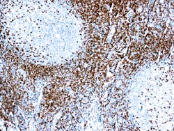
Diagnostic Biosystems CD138 CD138 (EP201)
CD138, also known as Syndecan-1, is a member of the transmembrane heparan sulfate proteoglycan family, acts as an extracellular matrix receptor and is involved in many cellular functions, including cell-cell adhesion and cell-matrix adhesion. CD138 expression is found in both hematopoietic and non-hematopoietic cells. In the hematopoietic system, CD138 labels plasma cells. It is an excellent marker for plasmacytic differentiation within the spectrum of hematologic malignancy. Among non-hematolymphoid cells, CD138 reactivity is observed in many types of epithelial cells and stoma cells in both normal and tumor tissues.
Diagnostic Biosystems CD1ACORTICALTHYMOCYTES CD1a Cortical Thymocytes
At least five CD1 genes (CD1a, b, c, d, and e) are identified. Cd1 proteins have been demonstrated to restrict T cell response to non-peptide lipid and glycolipid antigens and play a role in non-classical antigen presentation. CD1a is a non-polymorphic MHC Class I related cell surface glycoprotein, expressed in association with Beta-2 microglobulin. Anti-CD1a labels Langerhans cell histiocytosis (Histiocytosis X), extranodal histiocytic sarcoma, a subset of T-lymphoblastic lymphoma/leukemia, and interdigitating dendritic cell sarcoma of the lymph node. When combined with antibodies against TTF-1 and CD5, anti-CD1 is useful in distinguishing between pulmonary and thymic neoplasms since CD1a is consistently expressed in thymic lymphocytes in both typical and atypical thymomas, but only focally in 1/6 of thymic carcinomas and not in lymphocytes in pulmonary neoplasms. Anti-CD1a is reported to be a new marker for perivascular epithelial cell tumor (PEComa).

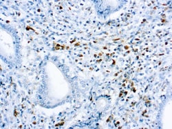
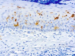
Diagnostic Biosystems MAST CELL TRYPTASE Mast Cell Tryptase
This antibody reacts with mast cells distributed in skin, synovium, lung, and heart. This antibody does not bind with any other cell type. Mast cells contain a number of preformed chemicals mediators such as histamine, chymase, carboxypeptidase, and proteolytic tryptase. Human mast cell tryptase is considered to be an important marker of mast cell activation and is an important mediator of inflammation.
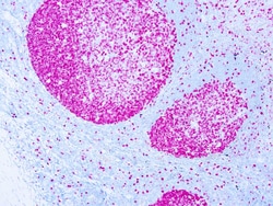
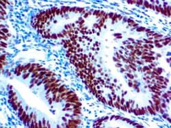
Diagnostic Biosystems KI-67 ANTIGEN Ki-67 Antigen
Ki-67 is a nuclear protein, which is expressed in proliferating cells. Ki-67 is preferentially expressed during late G1-, S-, M-, and G2-phases of the cell cycle, while cells in the G0 (quiescent) phase are negative for this protein.
Diagnostic Biosystems PLAP Placental Alkaline Phosphatase (PLAP) (SP15)
This antibody reacts with a membrane-bound isoenzyme (Regan and Nagao type) of placental alkaline phosphatase (PLAP) occurring in the placenta during the 3rd trimester of gestation. This antibody is highly specific to PLAP and shows no cross-reaction with other isoenzymes of alkaline phosphatases. It is useful in the identification of testicular germ cell tumors. Unlike germ cell tumors, PLAP-positive somatic cell tumors uniformly express epithelial membrane antigen (EMA).
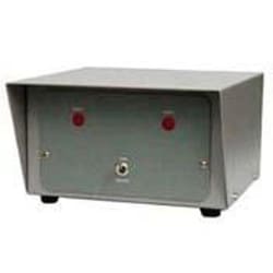
Bellco Glass, Inc. CO2 Tank Switch,115V
Provides automatic switching from the main tank to auxiliary tank.. Rear panel hose barbs are for gas connection. Alarm will sound when tank pressure drops below 5psi. Manual switching is done via the button labeled tank switch. Mounts on top of any stand. MADE IN THE USA