
Incubator Accessories

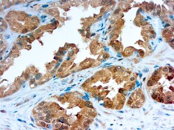
Diagnostic Biosystems GALECTIN-3 Galectin-3
Galectins are a family of soluble β-galactoside-binding animal lectins that modulate cell-to-cell adhesion and cell-to-extracellular matrix (ECM) interactions and play a role in tumor progression, pre-mRNA splicing and apoptosis. The galectin-3 protein, also known as Mac-2, hMac-2, GALBP, CBP35 or LGALS3, contains a single carbohydrate binding domain, which binds galactose-containing glycoconjugates. Galectin-3 is expressed in colonic and intestinal epithelium, inflammatory macrophages, papillary and follicular carcinomas, neoplastic astrocytes and some B and T lymphocytes. Upregulated expression of galectin-3 is involved in cancer progression and metastasis. Galectin-3 mediates the endocytosis of β1 Integrins in a lactose-dependent manner and is associated with thyroid malignancy and Crohn’s disease. It may also be used as a marker for diagnosing casesinvolving Hurthle cell adenomas and carcinomas.

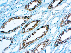
Diagnostic Biosystems RENAL CELL CARCINOMA Renal Cell Carcinoma (gp200)
This antibody recognizes a 200 kD glycoprotein (gp200) localized in the brush border of the proximal renal tebules and the luminal surface of Bowman’s capsule. gp200 was expressed by 93% of primary and 84% of metastatic renal carcinomas. Gp200 glycoprotein is also localized in the breast lobules and ducts, parathyroid glands and epididymis.
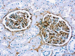
Diagnostic Biosystems FIBRONECTIN Fibronectin
This antibody reacts with human fibronectin in liver, tonsil, skin and kidney. Traces of contaminating antibodies have been removed by solid-phase adsorption. Only the fibronectin arch was observed in crossed immunoelectrophoresis.


Diagnostic Biosystems GLYCOPHORIN C Glycophorin C
This antibody is specific to β sialoglycophorin also known as glycophorin C. Glycophorin C is found on the human erythrocyte membrane and is one of four sialic acid-rich polypeptides.
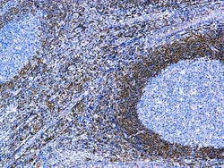
Diagnostic Biosystems BCL-2 ONCOPROTEIN Bcl-2 Oncoprotein
This antibody recognizes a protein of 25-26kDa, identified as bcl-2 alpha oncoprotein. It shows no cross-reaction with Bcl-x or Bax protein. Expression of bcl-2 alpha oncoprotein inhibits the programmed cell death (apoptosis). In most follicular lymphomas, neoplastic germinal centers express high levels of bcl-2 alpha protein, whereas the normal or hyperplastic germinal centers are negative. This antibody is valuable when distinguishing between reactive and neoplastic follicular proliferation in lymph node biopsies. It may also be used in distinguishing between those follicular lymphomas that express bcl-2 protein and small number in which neoplastic cells are bcl-2 negative.
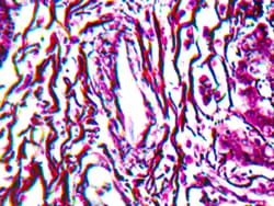
Diagnostic Biosystems RETICULUM STAIN KIT Reticulum Stain Kit
This kit stains reticulum fibers in paraffin tissue sections. The reticulum fibers stain gray to black, while the nuclei and background stain pink to rose.
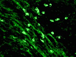
Diagnostic Biosystems IGM-FITC IgM-FITC
This antibody reacts with the Mu chain of human IgM. Traces of contaminating antibodies have been removed by solid phase absorption with human plasma proteins. This antibody is well suited for tissue section staining but may also be used for other applications.
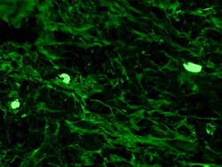
Diagnostic Biosystems IGG-FITC IgG-FITC
This antibody reacts with the gamma chain of human IgG. Traces of contaminating antibodies have been removed by solid phase absorption with human plasma proteins. This antibody is well suited for tissue section staining but may also be used for other applications.
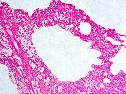
Diagnostic Biosystems PERMARED/AP PermaRed/AP
PermaRed/AP is a substrate-chromogen system designed to be used for either IHC or ISH when using alkaline phosphatase detection. PermaRed/AP produces a brilliant dark red color. PermaRed/AP is insoluble in organic solvents; therefore, sections can be dehydrated in alcohol, cleared in xylene (or a xylene-substitute), and permanently mounted. This chromogen substrate system may be used for both automation and manual use.
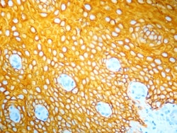
Diagnostic Biosystems PERMAYELLOW/HRP PermaYellow/HRP
PermaYellow/HRP is a substrate chromogen system for use with horseradish peroxidase (HRP) detection in immunohistochemistry (IHC) or in situ hybridizations (ISH). PermaYellow/HRP produces a distinct bright yellow that can easily be distinguished from other stains.
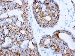
Diagnostic Biosystems E-CADHERIN E-Cadherin (SPM471)
Recognizes a protein of 120-80kDa, identified as E-cadherin. Cadherins comprise a family of Ca2+-depedent adhesion molecules that function to mediate cell-cell binding critical to the maintenance of tissue structure and morphogenesis. The c;assical cadherins, E-, N- and P-cadherin, consist of larg extracellular domains characterized by a series of five homologous NH2 terminal repeats. The relatively short intercellular domains interact with a variety of cytoplasmic proteins, such as β-catenin, to regulate cadherin function. E-cadherin plays an important role in epithelial cell adhesion. A decreased expression of E-cadherin is associated with metastatic potential and poor prognosis in breast cancer, prostate and esophageal cancer. In combination with p120 Catenin, it is useful for the differentiation between ductal (E-cadherin +) and lobular (E-cadherin -) breast carcinomas. It may also help in diagnosis of mesothelioma.
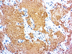
Diagnostic Biosystems PNEUMOCYSTIS CARINII Pneumocystis carinii
This antibody reacts with P. carinii in the fixed human tissues. The antibody is specific against an 82 kDa parasite specific polypeptide. It does not crossreact with G. lamblia, T. gondii, T. cruzi, L. tropica, E. histolytica, C. albicans and P. falciparum.
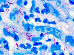
Diagnostic Biosystems FITE STAIN Fite's Stain
The Fite’s Stain Kit (For Leprosy and Nocardia) is intended for use in the histological visualization of mycobacterium Lepra bacillus and Nocardia. This kit may be used on formalin-fixed, paraffin-embedded sections.\n
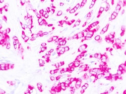
Diagnostic Biosystems EMA Epithelial Membrane Antigen (EMA) E29
This antibody reacts with an antigen of 265‐400 kDa belonging to a heterogeneous group of heavily glycosylated proteins called human milk fat globule proteins. It stains both normal and neoplastic cells. Among normal epithelia, it reacts strongly with mammary epithelium and glandular epithelia but shows a patchy staining with squamous epithelium.\n