
Primary Antibody Matched Pairs


Meso Scale Discovery RPLEX Mouse HAVCR1/KIM1 ASYQP

The R-PLEX Mouse Havcr1 (KIM-1) Assay by Meso Scale Diagnostics measures Havcr1 (KIM-1) in mouse serum plasma and cell culture supernatants It uses electrochemiluminescence (ECL) detection for high sensitivity a 5-log dynamic range and low sample volume The kit includes a 96-well plate with capture antibodies ECL-labeled detection antibodies and calibrator solutions for a standard curve The assay takes about 3 hours and is used to analyze Havcr1 as a biomarker of kidney toxicity and injury

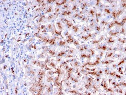
Biotium Cathepsin D (Tumor Marker)(CTSD/3276), Biotin conjugate, 0.1mg/mL
Cathepsin D is a ubiquitously expressed lysosomal aspartyl protease involved in the normal degradation of proteins. It is synthesized as an inactive 43 kDa preprocathepsin D that is cleaved and glycosylated to form a 46 kDa procathepsin D and then further cleaved to produce 28 kDa and 15 kDa subunits (heavy and light chains, respectively). Cathepsin D exhibits pepsin-like activity and plays a role in protein turnover and in the proteolytic activation of hormones and growth factors. Mutations in this gene play a causal role in neuronal ceroid lipofuscinosis-10 and may be involved in the pathogenesis of several other diseases, including breast cancer and possibly Alzheimer's disease. Primary antibodies are available purified, or with a selection of fluorescent CF Dyes and other labels. CF Dyes offer exceptional brightness and photostability. Note: Conjugates of blue fluorescent dyes like CF405S and CF405M are not recommended for detecting low abundance targets, because blu

Miltenyi Biotec, Inc. CD70 Antibody, anti-human, Vio Bright B515, REAfinity™
Identification and enumeration of CD70+ cells by flow cytometry.
This product is subject to the following: Miltenyi Biotec, Inc. Terms and Conditions of Sale For Products and Services

Sino Biological Anti-MEK2 Antibody, Rabbit Polyclonal 100 µL
Produced in rabbits immunized with a synthetic peptide corresponding to the center region of the Mouse MEK2, and purified by antigen affinity chromatography.
Genscript Corporation CD69/CLEC2C His Human
CLEC2C (CD69) is a membrane-bound type II C-lectin receptor and acts as a costimulatory molecule for T cell activation and proliferation It is involved in lymphocyte proliferation and functions as a signal transmitting receptor in lymphocytes natural killer (NK) cells and platelets CLEC2C is a disulfide-linked homodimer protein with two differentially glycosylated subunits
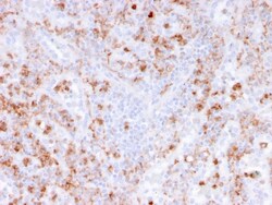
Biotium Calprotectin / MRP14 / S100A9 / Calgranulin B(rMAC3781), CF740 conjugate, 0.1mg/mL
Calprotectin is a heteroduplex of Protein S100-A8/A9 (also called MRP-8/14, or Calgranulin A/B). This antibody recognizes the 14 kDa B component (Protein S100-A9) of the complex. Protein S100-A9 is a member of the S100 family of proteins containing 2 EF-hand calcium-binding motifs. Altered expression of this protein is associated with the disease cystic fibrosis. This antibody reacts with neutrophils, monocytes, macrophages, and squamous mucosal epithelia and has been shown as an important marker for identifying macrophages in tissue sections. Primary antibodies are available purified, or with a selection of fluorescent CF Dyes and other labels. CF Dyes offer exceptional brightness and photostability. Note: Conjugates of blue fluorescent dyes like CF405S and CF405M are not recommended for detecting low abundance targets, because blue dyes have lower fluorescence and can give higher non-specific background than other dye colors.

Bioss ELAVL1 Recombinant Monoclonal
ELAVL1 Primary Antibody Unconjugated Host Species Rabbit Target Species Human Mouse Rat Clonality Monoclonal Applications WB FCM IHC-P IHC-F IF(ICC) IP IF RecombinantConcentration 1ug/ul Size 100 ul Storage Buffer 10mM phosphate buffered saline(pH 7 4) with 150mM sodium chloride 0 05% BSA 0 02% sodium azide and 50% glycerol
Miltenyi Biotec, Inc. MACSpep HiAg IFI30 (LLDVPTAAV) HLA-A*02
MACSpep single peptides developed for loading onto MHC multimers using the MHC MACSimer FleX Kits.
This product is subject to the following: Miltenyi Biotec, Inc. Terms and Conditions of Sale For Products and Services
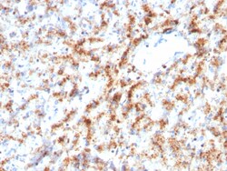
Biotium Cathepsin D (Tumor Marker)(CTSD/3082), CF488A conjugate, 0.1mg/mL
Cathepsin D is a ubiquitously expressed lysosomal aspartyl protease involved in the normal degradation of proteins. It is synthesized as an inactive 43 kDa preprocathepsin D that is cleaved and glycosylated to form a 46 kDa procathepsin D and then further cleaved to produce 28 kDa and 15 kDa subunits (heavy and light chains, respectively). Cathepsin D exhibits pepsin-like activity and plays a role in protein turnover and in the proteolytic activation of hormones and growth factors. Mutations in this gene play a causal role in neuronal ceroid lipofuscinosis-10 and may be involved in the pathogenesis of several other diseases, including breast cancer and possibly Alzheimer's disease. Primary antibodies are available purified, or with a selection of fluorescent CF Dyes and other labels. CF Dyes offer exceptional brightness and photostability. Note: Conjugates of blue fluorescent dyes like CF405S and CF405M are not recommended for detecting low abundance targets, because blu

Medchemexpress LLC Cellular retinoic acid-binding protein 2 (mouse), recombinant, His-tagged | >95.0% | 5 UG
Recombinant mouse cellular retinoic acid-binding protein 2 (CRABP2), N-terminal 6xHis-tagged, supplied lyophilized in a PBS-based buffer. The protein (partial sequence P2-E138) is produced in E. coli, has an approximate molecular weight of 16 kDa, and purity >95% by reducing SDS-PAGE. Supplied as 5 μg with recommended reconstitution concentration ≥100 μg/mL. Suitable for biochemical assays, binding studies, and antibody production.
- Expression system: E. coli
- N-terminal 6xHis tag for purification
- High purity greater than 95%
- Lyophilized from PBS, pH 7.4
- Supplied as 5 μg; reconstitute to ≥100 μg/mL
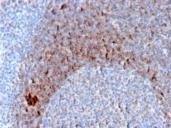
Biotium CD79b (B-Cell Marker)(B29/123), CF647 conjugate, 0.1mg/mL
CD79 (also designated Ig chains, designated CD79B or B29. The B cell antigen receptor complex (BCR) is formed by the association of CD79 with a membrane immunoglobulin, such as IgM or IgD. The membrane immunoglobulins IgM and IgD achieve surface expression and antigen presentation function in response to CD79 association. The cytoplasmic tails of both CD79A and CD79B contain an ITAM (immuno-receptor tyrosine-based activation) motif, which acts to initiate the BCR signaling reactions by binding to and activating tyrosine kinases. Primary antibodies are available purified, or with a selection of fluorescent CF Dyes and other labels. CF Dyes offer exceptional brightness and photostability. Note: Conjugates of blue fluorescent dyes like CF405S and CF405M are not recommended for detecting low abundance targets, because blue dyes have lower fluorescence and can give higher non-specific background than other dye colors.
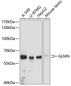
ABclonal Technology GLMN Rabbit pAb
This gene encodes a phosphorylated protein that is a member of a Skp1-Cullin-F-box-like complex. The protein is essential for normal development of the vasculature and mutations in this gene have been associated with glomuvenous malformations, also called glomangiomas. Multiple splice variants encoding different isoforms have been found for this gene.
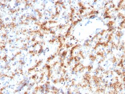
Biotium Cathepsin D (Tumor Marker)(CTSD/3082), CF740 conjugate, 0.1mg/mL
Cathepsin D is a ubiquitously expressed lysosomal aspartyl protease involved in the normal degradation of proteins. It is synthesized as an inactive 43 kDa preprocathepsin D that is cleaved and glycosylated to form a 46 kDa procathepsin D and then further cleaved to produce 28 kDa and 15 kDa subunits (heavy and light chains, respectively). Cathepsin D exhibits pepsin-like activity and plays a role in protein turnover and in the proteolytic activation of hormones and growth factors. Mutations in this gene play a causal role in neuronal ceroid lipofuscinosis-10 and may be involved in the pathogenesis of several other diseases, including breast cancer and possibly Alzheimer's disease. Primary antibodies are available purified, or with a selection of fluorescent CF Dyes and other labels. CF Dyes offer exceptional brightness and photostability. Note: Conjugates of blue fluorescent dyes like CF405S and CF405M are not recommended for detecting low abundance targets, because blu
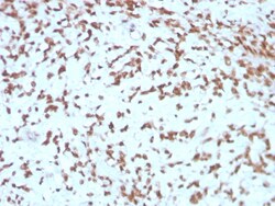
Biotium Cathepsin D (Tumor Marker)(CTSD/3275), CF488A conjugate, 0.1mg/mL
Cathepsin D is a ubiquitously expressed lysosomal aspartyl protease involved in the normal degradation of proteins. It is synthesized as an inactive 43 kDa preprocathepsin D that is cleaved and glycosylated to form a 46 kDa procathepsin D and then further cleaved to produce 28 kDa and 15 kDa subunits (heavy and light chains, respectively). Cathepsin D exhibits pepsin-like activity and plays a role in protein turnover and in the proteolytic activation of hormones and growth factors. Mutations in this gene play a causal role in neuronal ceroid lipofuscinosis-10 and may be involved in the pathogenesis of several other diseases, including breast cancer and possibly Alzheimer's disease. Primary antibodies are available purified, or with a selection of fluorescent CF Dyes and other labels. CF Dyes offer exceptional brightness and photostability. Note: Conjugates of blue fluorescent dyes like CF405S and CF405M are not recommended for detecting low abundance targets, because blu
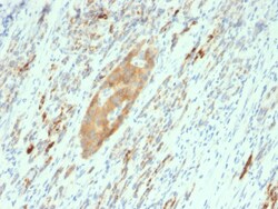
Biotium GAD2 / GAD65 (GABAergic Neuronal Marker)(GAD2/2362), CF405S conjugate, 0.1mg/mL
This MAb recognizes a protein of 65 kDa, which is identified as glutamic acid decarboxylase 2 (GDA2). It is responsible for catalyzing the production of gamma-aminobutyric acid from L-glutamic acid. There are two forms of glutamic acid decarboxylases (GAD's) that are found in the brain: GAD2 (also known as GAD65) and GAD1 (also known as GAD67). GAD1 and GAD2 are members of the group II decarboxylase family of proteins and are responsible for catalyzing the rate-limiting step in the production of GABA (-aminobutyric acid) from L-glutamic acid. Although both GAD's are found in the brain, GAD2 localizes to synaptic vesicle membranes in nerve terminals, while GAD1 is distributed throughout the cell. A pathogenic role for GAD2 is identified in the human pancreas since it has been identified as an autoantibody and an auto-reactive T cell target in insulin-dependent diabetes. Primary antibodies are available purified, or with a selection of fluorescent CF Dyes and other labels. CF Dyes