
Mouse Secondary Antibodies

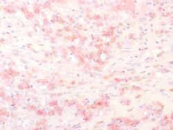
Biotium Tyrosinase-Related Protein-1 (TYRP-1) (Melanoma Marker), 1mg/mL
Reacts with a 75 kDa melanocyte-specific gene product, identified as Tyrosinase-related protein-1 (TRP-1). It is involved in melanin synthesis. TRP1 is present on the melanosomal membranes of melanoma, normal melanocytes and nevi. Recent evidence suggests that TRP-1 is involved in maintaining stability of tyrosinase protein and modulating its catalytic activity. TRP-1 is also involved in maintenance of melanosome ultrastructure and affects melanocyte proliferation and cell death. Primary antibodies are available purified, or with a selection of fluorescent CF Dyes and other labels. CF Dyes offer exceptional brightness and photostability. Note: Conjugates of blue fluorescent dyes like CF405S and CF405M are not recommended for detecting low abundance targets, because blue dyes have lower fluorescence and can give higher non-specific background than other dye colors.


Biotium CD257 / BAFF / TNFSF13B (C257/1638), CF740 conjugate, 0.1mg/mL
BAFF/CD257 is a type II transmembrane protein, and a member of the tumor necrosis factor ligand superfamily. It is proteolytically cleaved to form a soluble protein. BAFF, like other TNF ligand family members, forms homo-trimers. The predicted molecular weight of a BAFF monomer is approximately 31 kDa. It is expressed on peripheral blood B and T lymphocytes, monocytes, macrophages, and dendritic cells and is upregulated by IFN-gamma and down-regulated by PMA/ionomycin treatment. BAFF stimulates B and T cell immunity and regulates humoral immunity by binding TACI and BCMA receptors; BAFF prevents apoptosis and promotes B cell survival by binding the BAFF-specific receptor (BAFFR/BR3).Primary antibodies are available purified, or with a selection of fluorescent CF Dyes and other labels. CF Dyes offer exceptional brightness and photostability. Note: Conjugates of blue fluorescent dyes like CF405S and CF405M are not recommended for detecting low abundance targets, because

Biotium Cytokeratin 10 (Suprabasal Epithelial Marker) (DE-K10), CF488A conjugate, 0.1mg/mL
This MAb recognizes a protein of 56.5 kDa, identified as cytokeratin 10 (CK10). CK10 is expressed in all suprabasal layers of the epidermis. In the epidermis, expression of CK10 strictly parallels the extent of differentiation; it is absent in the basal layer, appears in the first suprabasal layers and increases in concentration towards the granular layer. However, CK10 is rarely detected in early stages of vulvar squamous carcinomas (tumors less than 2 cm, clinical stage I) regardless of the tumor grade. In larger and more advanced tumors (greater than 2 cm, clinical stages II and III), CK10 is detected very frequently. Expression of CK10 is related to maturation of malignant keratinocytes, being preferentially detected in more-differentiated parts.Primary antibodies are available purified, or with a selection of fluorescent CF Dyes and other labels. CF Dyes offer exceptional brightness and photostability. Note: Conjugates of blue fluorescent dyes like CF405S and CF405M

Biotium Erythropoietin (EPO) (EPO/1367), CF488A conjugate, 0.1mg/mL
This antibody recognizes a protein of about 37 kDa, which is identified as Erythropoietin (EPO). Erythropoietin is a secreted, glycosylated cytokine hormone composed of four alpha helical bundles. It is the primary factor responsible for regulating erythropoiesis during steady-state conditions and in response to blood loss and hemorrhage in the adult organism. Erythropoietin is synthesized by the kidney and stimulates the proliferation and maturation of bone marrow erythroid precursor cells. The protein is found in the plasma and regulates red cell production by promoting erythroid differentiation and initiating hemoglobin synthesis.Primary antibodies are available purified, or with a selection of fluorescent CF Dyes and other labels. CF Dyes offer exceptional brightness and photostability. Note: Conjugates of blue fluorescent dyes like CF405S and CF405M are not recommended for detecting low abundance targets, because blue dyes have lower fluorescence and can give higher
Biotium Neurofilament (NF-L) (Neuronal Marker) (NEFL/2983R), CF740 conjugate, 0.1mg/mL
This MAb reacts with a 68 kDa protein, identified as light sub-unit of neurofilaments (NF-L). Neurofilaments make up the main structural elements of axons and dendrites and are found in neurons, peripheral nerves, and sympathetic ganglion cells. Neurofilaments consist of three major subunits with molecular weights of 68 kDa (NF-L), 160 kDa (NF-M) and 200 kDa (NF-H). Anti-neurofilament stains a number of neural, neuroendocrine, and endocrine tumors. Neuromas, ganglioneuromas, gangliogliomas, ganglioneuroblastomas, and neuroblastomas stain positively for anti-neurofilament. Neurofilaments are also present in paragangliomas as well as adrenal and extra-adrenal pheochromocytomas. Carcinoids, neuroendocrine carcinomas of the skin, and oat cell carcinomas of the lung also express neurofilament.Primary antibodies are available purified, or with a selection of fluorescent CF Dyes and other labels. CF Dyes offer exceptional brightness and photostability. Note: Conjugates of blue fluores
Biotium CDX2 (GI Epithelial Marker) (CDX2/2951R), CF740 conjugate, 0.1mg/mL
The intestine-specific transcription factors CDX1 and CDX2 are important for directing intestinal development, differentiation, proliferation and maintenance of the intestinal phenotype. CDX2 protein expression has been seen in GI carcinomas. Anti-CDX2 has been useful to establish GI origin of metastatic adenocarcinomas and carcinoids and is especially useful to distinguish metastatic colorectal adenocarcinoma from lung adenocarcinoma. However, mucinous carcinomas of the ovary also express CDX2 protein. It limits the usefulness of this marker in the distinction of metastatic colorectal adenocarcinoma from mucinous carcinoma of the ovary.Primary antibodies are available purified, or with a selection of fluorescent CF Dyes and other labels. CF Dyes offer exceptional brightness and photostability. Note: Conjugates of blue fluorescent dyes like CF405S and CF405M are not recommended for detecting low abundance targets, because blue dyes have lower fluorescence and can give higher

Meridian Life Science MAb to Canine NT-proBNP
pro-Brain Natriuretic Peptide, N-Terminal (NT-proBNP) (a.a. 40-50)
Immunology Consultants anti-Dog IgE Antibody (Mouse - Clone #E671) - Monoclonal
This monoclonal antibody targets a distinct canine IgE epitope, providing sensitive, specific detection in ELISA and Western blotting. Clone E671 offers strong affinity, low background, and consistent lot‑to‑lot performance. It is well suited for veterinary immunology research, allergy diagnostics, and studies examining IgE‑mediated immune responses in dogs. ME‑40A‑E671 delivers reliable, high‑clarity detection across varied sample types.
Immunology Consultants anti-Dog CRP Antibody (Mouse - Clone #14A2) - Monoclonal
This monoclonal antibody provides sensitive, specific detection of canine C‑reactive protein (CRP), a major acute‑phase biomarker. Clone 14A2 performs reliably in ELISA, Western blotting, and antigen‑capture assays, offering strong affinity and low background. It is ideal for veterinary diagnostics, inflammation research, and monitoring CRP levels in canine serum or plasma. MCRP‑40A‑14A2 delivers consistent, high‑clarity performance across repeated assays.
Vector Laboratories Monoclonal Mouse Anti‑Dog IgG Secondary Antibody (clone DIG12‑255.5) LS‑C152977, 100ug
Dog IgG antibody LS-C152977 is an unconjugated mouse monoclonal antibody. Validated for ELISA.

Vector Laboratories Monoclonal Mouse Anti‑Dog IgG Secondary Antibody (clone DIG11‑127.2) LS‑C760090, 100ug
Dog IgG antibody LS-C760090 is an unconjugated mouse monoclonal antibody. Validated for ELISA.
Immunology Consultants anti-Dog IgE Antibody (Mouse - Clone #2A2) - Monoclonal
This monoclonal antibody provides high‑specificity detection of canine IgE, supporting ELISA, Western blotting, and allergy‑focused assay development. Clone 2A2 delivers strong affinity and low background, enabling sensitive measurement of IgE‑mediated responses in dogs. It is well suited for veterinary diagnostics, hypersensitivity research, and studies examining allergic disease progression. ME‑40A‑2A2 offers consistent, high‑clarity performance across repeated assays and complex biological samples.
Immunology Consultants Monoclonal Mouse anti-Dog IgM Antibody Clone 3H6, 1.0 mg
This mouse IgG2a is purified using Protein A chromatography. It is produced from tissue culture media obtained at our facility in The United States. Antibody concentration was determined using an absorbance at 280 nm: 1.4 equals 1.0 mg of IgG. This antibody reacts with dog IgM as determined by ELISA and western blotting. No cross-reactivity was detected with dog IgG and IgA by indirect ELISA. Optimal working dilutions should be determined experimentally by the investigator.