
Histology Reagents


Electron Microscopy Sciences 10% Glutaraldehyde in Solvent Of Choice 10 ML
C₅H₈O₂
Rapid-Freezing on a polished metal surface, at the temp. of liquid helium or liquid nitrogen, followed by freeze-substitution in osmium-acetone, has been shown to be a very useful technique for capturing rapid cellular events and for preserving cell ultrastructure.

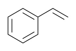
Electron Microscopy Sciences Styrene, Reagent 450 ML
(Vinylbenzene)
CAS #100-42-5 C₆H₅CH:CH₂ F.W. 104.14
Assay: >99.0%
A monomeric styrene used as an embedding media for ultramicrotomy. (DeLamater, E.D., et al 1971). A styrene-methacrylate resin embedding method for ultrathin sectioning ( J. Electron Micro., 10:16)

Electron Microscopy Sciences Acid-Fuchsin Aniline Solution 500 ML
Used in Laqueur's Method for Alcoholic Hyalin for mallory bodies, erythrocytes, bile pigment and proteinaceous, material in liver.
Laqueur, G.I., Amer. J. Clin. Path., 20:689-690, 1950, Luna, L.G. (ed), Manual of Hitopathologic Staining Method of the AFIP, 3rd ed, McGraw-Hill, NY, c. 1968, p 160
Lillie, R.D., Fullmer, H.M., Histopathologic Technic and Practical Histochemistry 4th ed., cGraw-Hill, NY, 1976, p 670-671
Clark, G., (ed) Staining Procedures, 3rd ed. WilliamS & Wilkins, Baltimore, p 51, c. 1973
Biocare Medical, LLC XM Factor - 1.0 ml
The XM Factor is a potent blocker for eliminating endogenous mouse IgG in mouse tissues. It has been specially designed to work with Biocare Rabbit-on-Rodent and Goat Polymer detection systems. XM Factor cannot be used with Mouse-on-Mouse AP or HRP polymer. By adding XM Factor to the polymer detection, endogenous mouse IgG is dramatically reduced and/or is completely eliminated. Biocare has also developed Rodent Block M that blocks for nonspecific background staining, and at the same time, also helps reduce endogenous mouse IgG. The combination of Rodent Block M and the XM Factor provide clean staining for mouse tissue protocols. It can also be used on an automated staining system.
Electron Microscopy Sciences Acid Alcohol 1% 500 ML
References: Genta PM., Robason GO, Graham DY-Simultaneous Visualization of Helicobactor Pylon and Gastric Morphology A New Stain Hum Pathol. 25:22 1-226, 1994.
Crescent Chemical Co Inc GLUTARALDEHYDE 25%
Glutaraldehyde 25 % solution in water for electron microscopy, high purity In vials; filled under argon. Size - 10x5ML Storage Conditions - +2 °C TO +8 °C Catalog Number - 23114.02

Electron Microscopy Sciences Acetate Buffer, 0.2m, pH 5.0 500 ML
Acetate Buffer in 0.2M solution with a pH of 5.0.
Electron Microscopy Sciences Collodion (Parlodion), 2% in Amyl Acetate 30 ML
Highly purified nitrocellulose (parlodion strip) in glass distilled amyl acetate. Useful for forming a negative replica to very fine detail. Two types are available: Our sterile formula which is filtered down to 0.45 micron and our non-sterile formula. Non-Filter 2% Collodion. 30 ml container avoids hazardous shipping.
Electron Microscopy Sciences Collodion (Parlodion), in Amyl Acetate 30 ML
Highly purified nitrocellulose (parlodion strip) in glass distilled amyl acetate. Useful for forming a negative replica to very fine detail. Two types are available: Our sterile formula which is filtered down to 0.45 micron and our non-sterile formula. Sterile formula is filtered to 0.45 micron. 30 ml avoids hazardous shipping.

Electron Microscopy Sciences Eosin Y, Certified, C.I. DcE-40 10 GR
(Acid Red 87, Eosin Yellowish)
CAS #17372-87-1 C₂cH₆Br₄Na₂O₅ F.W. 691.88
Solubility: 44.0% Water; 2.0% Alcohol; 25.0% Cellosolve; 27.5% Glycol; 0.0% Xylene
Eosin Y is used with Hematoxylin for showing cell nuclei in embryos and kidneys (Conn, "Biological Stain" 9th ed., The Williams and Wilkens Co., Baltimore 1977), as a component of Wright's stain for blood corpuscles (Wright, J. Med. Res., 7, 138 (1902), and for staining bone marrow to show cell morphology (Bech, "Lab. Manual of Hematological Tech.", W. B. Saunders Co., Phila., 1908, p. 233).
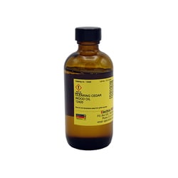
Electron Microscopy Sciences Clearing Cedar Wood Oil 100 ML
Refractive Index 20 degree C 1.504+/-.002. For clearing.
Electron Microscopy Sciences Acetate Buffer, 0.2m, pH 5.6 500 ML
Acetate Buffer in 0.2M solution with a pH of 5.6.
Electron Microscopy Sciences Amphibian Ringer Solution 1 LT
Amphibian Ringer Solution is made up of salts including sodium chloride, potassium chloride, and calcium chloride hexahydrate.
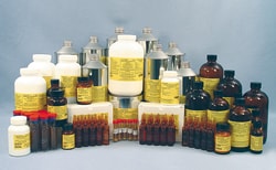
Electron Microscopy Sciences Chemkit for Preparations of Biological Specimens
This package is arranged to enable a laboratory to perform standard preparations of biological specimens with enough supplies to last. Includes:
Fixatives-osmium tetroxide crystal, osmium tetroxide 4% solution, glutaraldehyde EM grade 50%, glutaraldehyde EM grade 8%, and paraformaldehyde Prill.
Buffers-sodium phosphate monobasic, sodium phosphate dibasic, sodium hydroxide pellets, and sodium cacodylate trihydrate.
Dehydrants-propylene oxide and acetone.
Embedding media-EMBed-812 kit and low viscosity kit (Dr. Spurr).
Stains-toluidine blue, borax tetra, sodium borate, uranyl acetate, lead citrate, and epoxy tissue stain.
Visikol Inc Visikol HISTO 1 & 2 Combo (100 mL each)

Laboratory Reagent, Histological Reagent, Tissue Clearing Reagent, Visikol HISTO-1 and HISTO-2 Combo, 100mL each. A simple and rapid tissue clearing formula that can be used to image tissues in 3D without damaging the underlying cellular morphology. Designed to pair with immunolabeling and reversible so that 3D histology can be followed up with traditional 2D histology. The Visikol HISTO tissue clearing approach is comprised of two different reagents called Visikol HISTO-1 and Visikol HISTO-2 whereas Visikol HISTO-1 is used first and Visikol HISTO-2 is used second. However, for small tissues such as micro-tissues and thin brain slices (500 um), only Visikol HISTO-1 is required and tissues can be imaged in Visikol HISTO-1. Larger tissues must be placed in Visikol HISTO-2 to finish clearing and then these samples can be imaged in Visikol HISTO-2. WARNING: Do not use HISTO-2 within any polystyrene vessle.