
Incubator Accessories


Diagnostic Biosystems ADRENOCORTICOTROPIC HORMONE Adrenocorticotropic Hormone (ACTH) - (AH26)
This antibody is specific to Synacthen (aa1-24 of ACTH); does not react with CLIP (aa17-39 of ACTH). POMC (peo-opiomelanocortin or corticotropinlipotropin) is a 267 amino acid polypeptide hormone precursor that goes through extensive, tissue-specific posttranslational processing by convertases. This antibody stains corticotrophs in the adenohypophysis and is useful in the classification of pituitary adenomas.


Diagnostic Biosystems OCT 3/4 OCT-3/4 (OCT4/6847R)
OCT-3/4 (also known as POU5F1) is a transcription factor that has been recognized as fundamental in the maintenance of pluripotency in embryonic stem cells and primordial germ cells. It has been proposed as a useful marker for germ cell tumors (GCT) that exhibit features of pluripotentiality (seminoma/dysgerminoma, germinoma and embryonal carcinoma. OCT-3/4 immunostaining has been shown to be sensitive and specific for GCT, whether in primary gonadal or extragonadal sites or in metastatic lesions.

Diagnostic Biosystems DP3 DP3 for One-Step Deparaffinization/20 Runs
DP3 is a blend of aqueous buffers and a paraffin solvent (orange oil). Because of these properties it can act as both a deparaffinzing agent and a reydration agent thus eliminating the requirement for transferring the paraffin tissues through a series of graded xylenes and alcohol. Deparaffinization occurs rapidly at temperatures in excess of 50 C. (Range 50 - 80 C). In contrast to other aqueous deparaffinization reagents, the paraffin is completely removed from both around and beneath the tissues.\n\nDP3 deparaffinization is compatible with Immunohistochemistry.
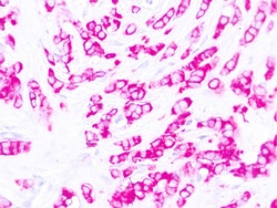
Diagnostic Biosystems EMA Epithelial Membrane Antigen (EMA) E29
This antibody reacts with an antigen of 265‐400 kDa belonging to a heterogeneous group of heavily glycosylated proteins called human milk fat globule proteins. It stains both normal and neoplastic cells. Among normal epithelia, it reacts strongly with mammary epithelium and glandular epithelia but shows a patchy staining with squamous epithelium.\n

Diagnostic Biosystems UROPLAKIN 11 UPK2 Uroplakin II
Uroplakins (UPs) are a family of transmembrane proteins (UPs Ia, Ib, II and III) that are specific differentiation products of urothelial cells. Uroplakins are markers of terminally differentiated urothelium. Uroplakin II (UPII) is a newly described sensitive marker for urothelial carcinoma (UC). The expression profile of UPII in different types of UC and its utility in the diagnostic setting are needed.
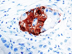
Diagnostic Biosystems SYNAPTOPHYSIN SYP02 Synaptophysin (SYP02)
This antibody isspecific to a 38 kDa protein. Synaptophysin is an glycoprotein occurring in presynaptic vesicles of neurons in the brain, spinal cord, retina, vesicles of adrenal medulla and in neuromuscular junctions. This antibody reacts with neuroendocrine neoplasms of neural as well as epithelial types.

Diagnostic Biosystems GLYCOPHORIN C Glycophorin C
This antibody is specific to β sialoglycophorin also known as glycophorin C. Glycophorin C is found on the human erythrocyte membrane and is one of four sialic acid-rich polypeptides.
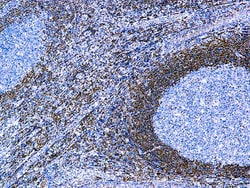
Diagnostic Biosystems BCL-2 ONCOPROTEIN Bcl-2 Oncoprotein
This antibody recognizes a protein of 25-26kDa, identified as bcl-2 alpha oncoprotein. It shows no cross-reaction with Bcl-x or Bax protein. Expression of bcl-2 alpha oncoprotein inhibits the programmed cell death (apoptosis). In most follicular lymphomas, neoplastic germinal centers express high levels of bcl-2 alpha protein, whereas the normal or hyperplastic germinal centers are negative. This antibody is valuable when distinguishing between reactive and neoplastic follicular proliferation in lymph node biopsies. It may also be used in distinguishing between those follicular lymphomas that express bcl-2 protein and small number in which neoplastic cells are bcl-2 negative.

Diagnostic Biosystems LBA1 Iba1 (AIF1)
Actin-binding protein that enhances membrane ruffling and RAC activation. Enhances the actin-bundling activity of LCP1. Binds calcium. Plays a role in RAC signaling and in phagocytosis. AIF1 colocalizes with actin, and upon stimulation, translocates to lamellipodia. It is also a marker of human microglia and is expressed by macrophages in injured skeletal muscle. The gene encoding AIF1 resides in the tumor necrosis factor (TNF) cluster of genes, located in the region represented by the human major histocompatibility complex (MHC).\n
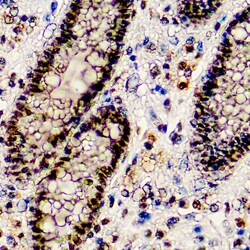
Diagnostic Biosystems BAP1 BAP1 (BAP1/2667)
BAP1 (BRCA1-Associated Protein 1) was initially identified as a protein that binds to BRCA1. BAP1 is a tumor suppressor that is believed to mediate its effects through chromatin modulation, transcriptional regulation, and possibly via the ubiquitin-proteasome system and the DNA damage response pathway. Germline mutations of BAP1 confer increased susceptibility for the development of several tumors, including uveal melanoma, epithelioid atypical Spitz tumors, cutaneous melanoma, and mesothelioma. Somatic BAP1 mutations are seen in cutaneous melanocytic tumors (epithelioid atypical Spitztumors and melanoma), uveal melanoma, mesothelioma, clear cell renal cell carcinoma, and other tumors. BAP1 immunohistochemistry is particularly useful in differentiating malignant mesothelioma (nuclear negative) vs. reactive mesothelial proliferation (nuclear positive).

Diagnostic Biosystems CADHERIN 17 Cadherin 17
Cadherin 17 is exclusively expressed in the epithelial cells of embryonic and adult small intestine, colon, liver, and pancreatic ducts. It has been reported to be frequently expressed in adenocarcinomas arising in the gastrointestinal tract and pancreas. Owing to its restricted expression in these groups of tumors, cadherin 17 has proven to be a useful immunohistochemical marker for assisting in distinguishing these neoplasms from other malignancies with which they may be confused

Diagnostic Biosystems P27 KIP1 p27KIP1, Mitotic Inhibitor / Suppressor Protein
This antibody is specific to a protein of 27 kDa known as p27Kip1, a cell cycle regulatory mitotic inhibitor. Its epitope spans between aa 83-204 of p27. It is highly specific and shows no cross-reaction with other related mitotic inhibitors. p27Kip1 functions as a negative regulator of G1 progression and has been proposed to function as a possible mediator of TGF- induced G1 arrest. p27Kip1 is a candidate tumor suppressor gene. Down regulation of p27Kip1 is found in many types of cancer, and decreased expression of p27Kip1 is associated with poor prognosis in several tumors, including lung, breast, colorectal and prostate.
Diagnostic Biosystems CD7 CD7 (CBC.37.80)
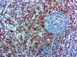
CD7 is expressed by most peripheral blood T cells, NK cells, and all thymocytes. It is one of the earliest surface antigens on T and NK-cell lineages. The antibody is a useful aid for classification of T-cell malignancies.

Diagnostic Biosystems HEMATOXYLIN Montage™ Hematoxylin
Hematoxylin is a histological staining reagent suitable for visualization of nuclei in tissue sections. Montage™ Hematoxylin, is designed for optimal performance when used together with the Montage 360™ System.\n\nDBS’s Hematoxylin is intended for use in the histologic demonstration of nuclear staining. This staining technique is used to make the critical distinction between a normal nucleus and an abnormal one.

Diagnostic Biosystems CD7 CD7 (CBC.37.80)
CD7 is expressed by most peripheral blood T cells, NK cells, and all thymocytes. It is one of the earliest surface antigens on T and NK-cell lineages. The antibody is a useful aid for classification of T-cell malignancies.