
Cellular Imaging Instrumentation
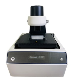
LAXCO Accuva LEAP upgradable multichannel phase-fluorescence inverted imaging system

Motorized X-Y-Z stage. Eyepieceless design.
LAXCO Accuva LEAP Upgradable Multichannel Phase-Fluorescence Inverted Imaging System PROMO
Motorized X-Y-Z stage. Eyepieceless design.

LAXCO Accuva LEAP Upgradable Phase Inverted Imaging System PROMO
Motorized X-Y-Z stage. Eyepieceless design.
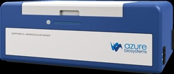
Azure Biosystems Sapphire FL Biomolecular Imager
Provides upgradable laser scanner with a wide field of view, selection of user-adjustable lasers and filters

Thermo Scientific™ CellInsight™ CX5 High Content Screening (HCS) Platform
Rapidly perform high content imaging assays with this affordable, compact HCS platform.


Thermo Scientific™ CellInsight™ CX7 LZR High Content Analysis Platform and Store Standard Edition (SE)
CellInsight CX7 LZR High Content Analysis Platform and Store Standard Edition (SE)

Thermo Scientific™ CellInsight™ CX5 High Content Screening (HCS) Platform
Rapidly perform high content imaging assays with this affordable, compact HCS platform.
Thermo Scientific™ Onstage Incubator for CellInsight CX5/NXT HCS Platforms
Onstage Incubator for CellInsight CX5/NXT HCS Platforms
Thermo Scientific™ CellInsight™ CX7 LZR High Content Analysis Platform
Thermo Scientific™ CellInsight™ CX7 LZR High Content Analysis Platform is fast, laser-based, and designed for quantitative microscopy.

Thermo Scientific™ Onstage Incubator for CellInsight CX7 HCA Platform
Onstage Incubator for CellInsight CX7 HCA Platform
Sartorius INCUCYTE S3 HD/2CLR SYSTEM
The Incucyte S3 Live-Cell Analysis System is a high-performance instrument designed to image and analyze living cells around the clock for days weeks or months while cells remain undisturbed inside a standard tissue culture incubator. Incucyte S3 is designed for handling higher throughput and accommodates up to six microplates at a time while imaging at 4X 10X or 20X. The Incucyte S3 is a two-color system that comes standard with a Green/Red Optical Module Orange/NIR Optical Module sold separately with HD phase contrast imaging that supports a wide range of 2D and 3D cell culture applications. Multi-user support allows scheduling of experiments at different image acquisition frequencies and magnifications in parallel.

Yamato Scientific America, Inc. Cell Imager BF + Green Fluo 4x
Celloger Mini Plus is an automated live cell imaging system that is equipped with an advanced fluorescence and bright field microscopy, autofocusing and real time multi-position imaging technology. Allows easy monitoring of live cells inside the incubator for a long time without disturbing the environment suitable for cell culture. It doesn’t have a moveable stage but instead, the camera inside the system moves to capture the images of cell in multiple positions. Since the vessel and cell sample are in a steady state, this provides a stable environment for the cells to grow. Its compact size can fit into a standard CO2 incubator and can easily be carried in and out of the incubator during the experiment.
RWD LIFE SCIENCE INC C100 Automated Cell Counter C100-Pro, Bright Field and Flourencent Field, Cell Counting and Analysis in 9s
A Real Time-Saver Speeds up Your Research.RWD automated cell counters provide two models to meet different customers’ needs in BF and FL cell counting and analysis in 9s. Combined with highly intelligent software and excellent microscopy optics structure, the counters could liberate researchers from the heavy work of daily cell counting